


Structural Proteomics
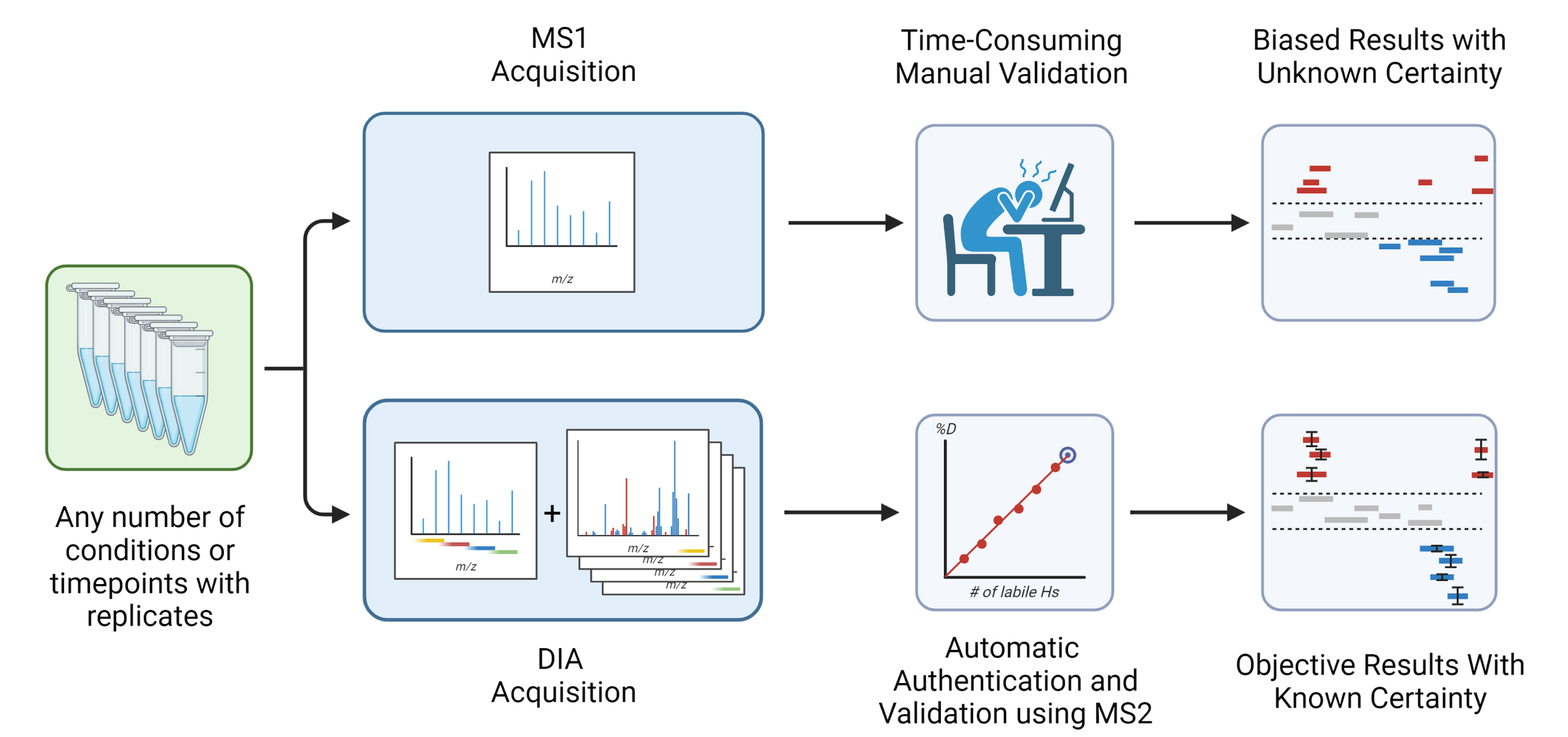
Hydrogen-deuterium exchange is a structural method that monitors the dynamics of proteins. The method supports the analysis of protein-protein and protein-drug interactions and the measurement of protein stability. But it could be so much more if the technology was more sensitive, complexity tolerant and faster. Tandem MS provides this capability. We are advancing some exciting technology and software to make this a high-throughput ultrasensitive assay.
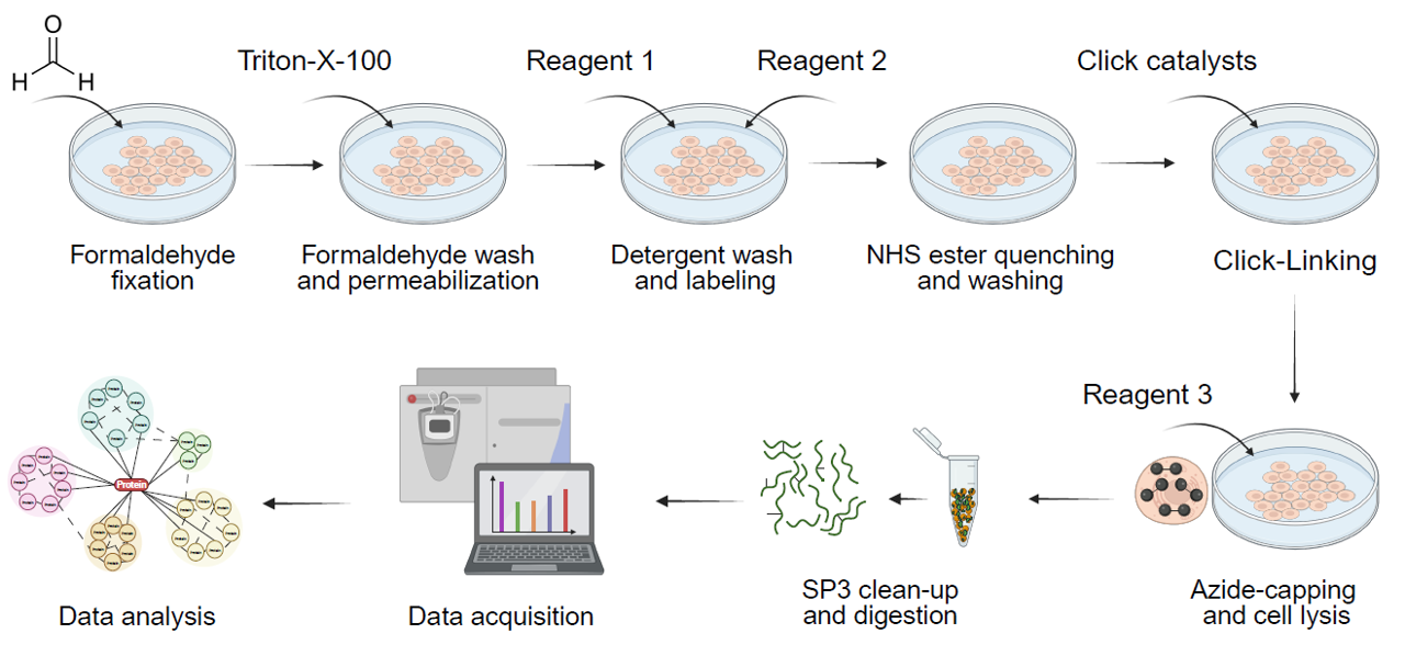
Crosslinkers are “molecular rulers” that can return actual distance measurements. If we can install these crosslinkers in cells at high yield we can vastly improve our measurements of the cellular interactome. We figured out how. However, detecting them is another matter. Our research program in this area focuses on how to improve crosslink identification by mass spec, through new reagents, methods and software.
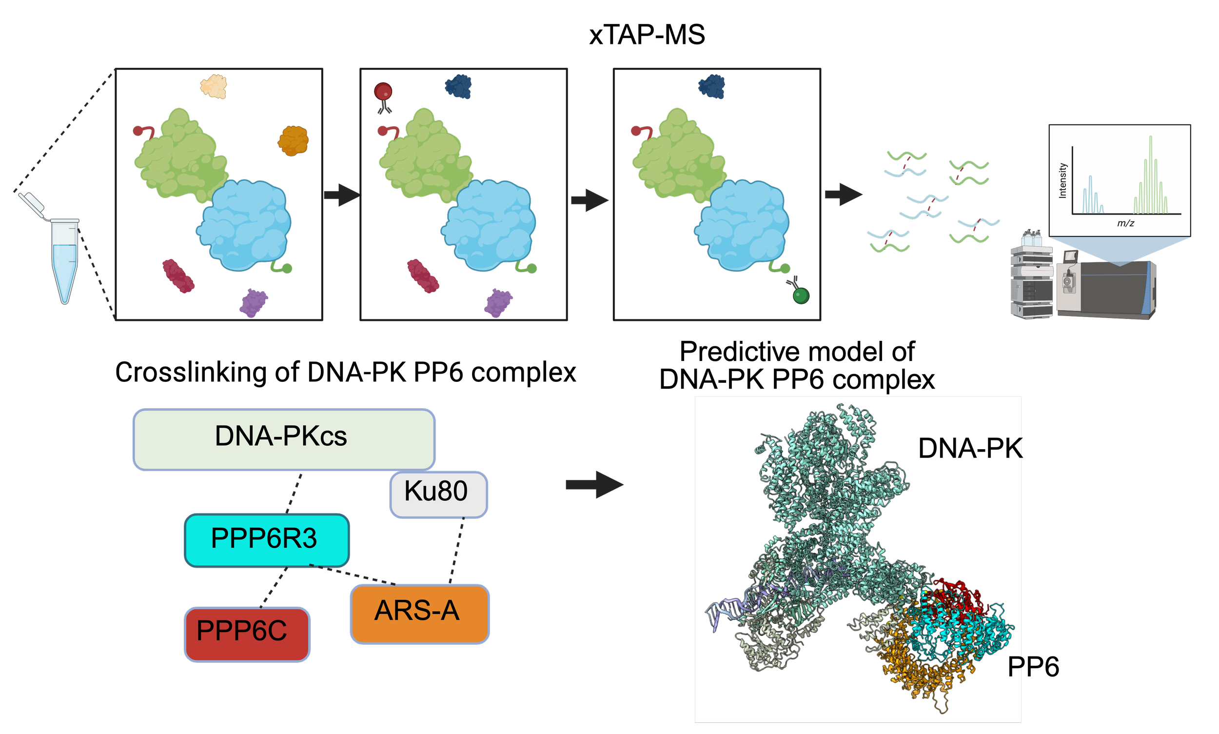
xTAP-MS
Our latest crosslinking method. It should be possible to detect crosslinks for your complex of interest and support integrative modeling. Unfortunately, direct affinity isolation rarely works. Enter xTAP-MS, a method for detecting abundant crosslinks on targeted complexes. It works because it addresses the signal splitting phenomenon, but we still need to turn the strategy in to a universal and scalable solution.